
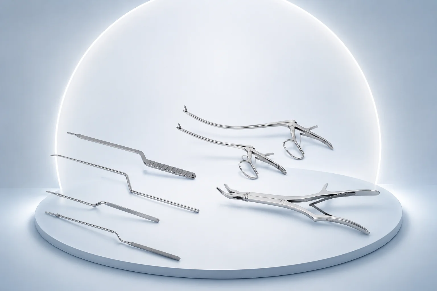
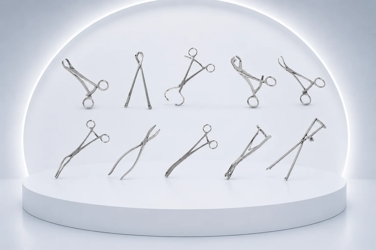
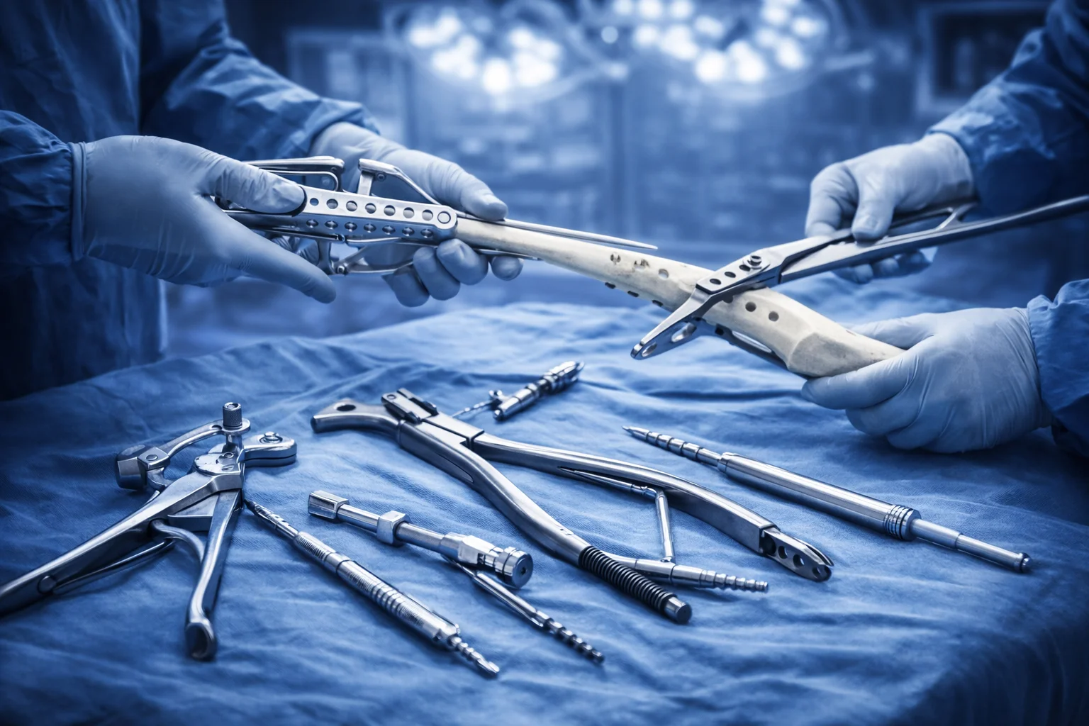
Employed to compress and align the anterior pelvic ring during symphyseal plating and acetabular fracture approaches through the ilioinguinal incision. The caliper jaws span the pelvic brim to reduce diastasis of the pubic symphysis before screw fixation. Pelvic surgeons use the integrated measurement scale to confirm anatomic reduction against preoperative templating.
Used in the operating room to flush-cut Kirschner wires and cerclage wire after passage through bone tunnels in open reduction internal fixation. The tungsten carbide inserts deliver clean shear cuts that reduce wire burrs at the cortical surface of the tibia and femur. Orthopedic teams rely on the double-action leverage to cut 1.6–2.0 mm pins without deforming adjacent soft tissue.
Used to seat and advance spinal or long-bone fixation rods into pedicle screw tulips and external fixator clamps during construct assembly. The pistol-grip handle directs axial force along the rod axis when contouring rods to match lumbar lordosis. Spine surgeons employ it to reduce spondylolisthesis by pushing the rod into the screw heads before set screw tightening.
Employed during trauma fixation to trim Steinmann pins and tension-band wires protruding from the olecranon or patella after fracture stabilization. The full tungsten carbide jaws maintain sharpness through repeated cuts on hardened stainless fixation wire during long ORIF cases. Surgeons use the compound hinge to generate cutting force with minimal hand fatigue when multiple pins require trimming at case conclusion.
Contours reconstruction plates to match the anatomy of the clavicle, distal radius, and proximal humerus before intraoperative application. The set includes pliers for in-plane and out-of-plane bending so the plate sits flush against the bone surface without gapping. Orthopedic technicians and surgeons use it during ORIF when pre-contoured plates require final adjustment at the fracture site.
Applied to resect thickened bone edges, osteophytes, and small bone fragments during joint debridement and fracture site preparation. The heavy-duty double-action mechanism provides the mechanical advantage needed to nibble cortical bone in revision arthroplasty and nonunion takedown. Orthopedic surgeons use it to contour bone surfaces before plate application on comminuted diaphyseal fractures.
Grasps fracture fragments at an oblique angle during reduction of metadiaphyseal tibia and distal radius fractures where straight forceps cannot achieve perpendicular purchase. The speed-lock ratchet maintains compression across the fracture while the surgeon drills and inserts lag screws. Its angled jaws facilitate reduction in confined wound corridors created by minimally invasive plate osteosynthesis.
Used to contour and insert cerclage or tension-band wire through pre-drilled holes in the proximal humerus and distal radius during fracture fixation. The pliers bend wire to match the bone contour before the surgeon seats the loop and twists the free ends for compression. They are essential when constructing figure-of-eight wire patterns around the olecranon or medial malleolus.
Temporarily clamps bone segments or plate-bone constructs during complex periarticular fracture fixation when multiplanar alignment must be held. The articulated joint allows the surgeon to adjust the clamp angle to match the fracture obliquity at the distal femur or proximal tibia. It serves as a third hand while definitive screws are placed through a locking compression plate.
Grasps long bone fragments during plating of femoral and tibial shaft fractures while the threaded locking mechanism maintains compression across the fracture site. The integrated thread allows incremental tightening without releasing grip on osteoporotic bone. Surgeons use these forceps to hold reduction while drilling screw holes and applying the definitive reconstruction plate.