
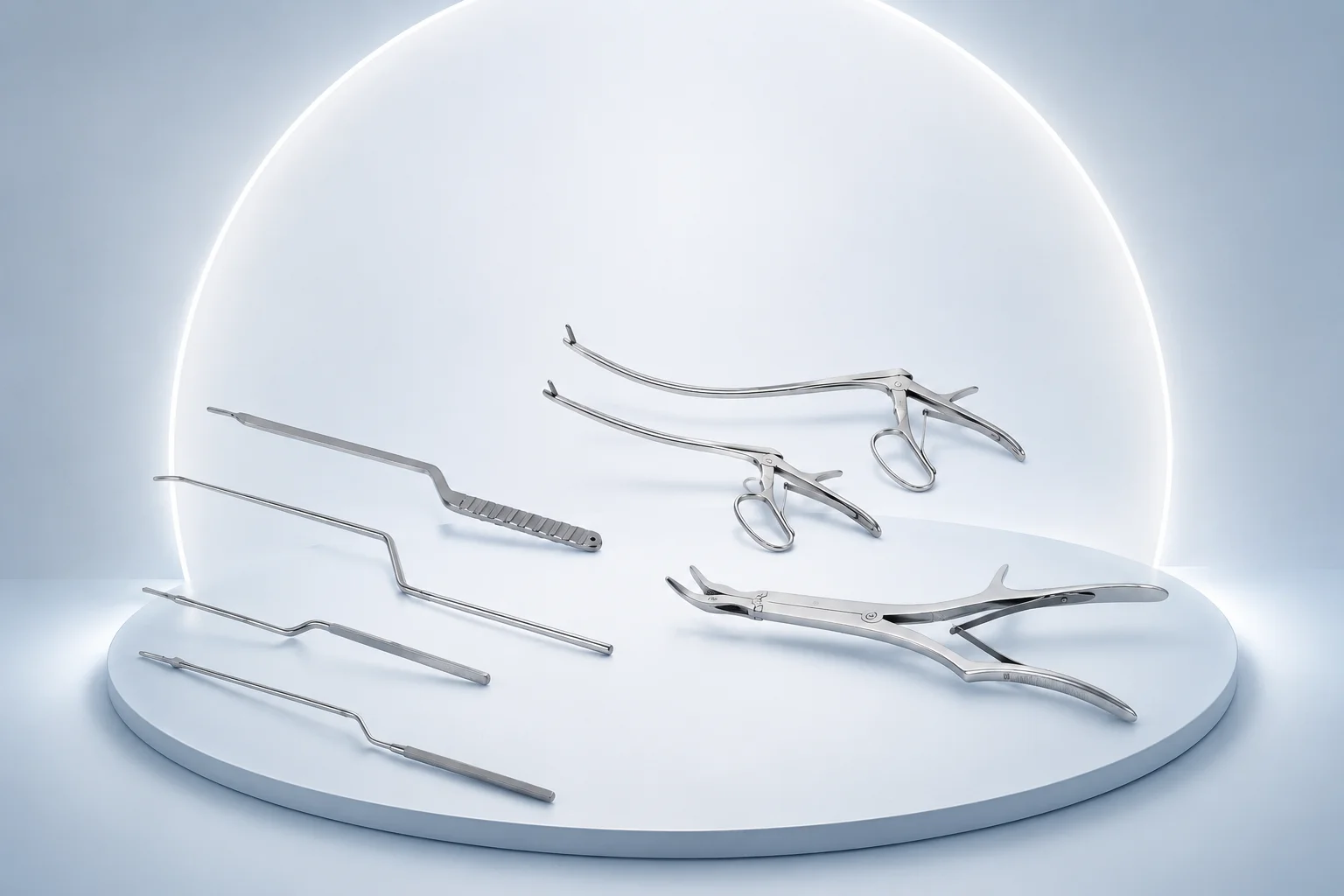
Ball-tip probes navigate the medullary canal and fracture site to assess cortical continuity before reaming for intramedullary nailing of femoral shaft fractures. The smooth ball tip prevents perforation of the anterior femoral cortex during antegrade nailing through the greater trochanter. Trauma surgeons advance the probe to confirm canal patency after guidewire placement.
Scrapes cancellous bone and fibrous tissue from the fracture bed of the tibial plateau and distal radius during open reduction to promote healing. The angled shaft reaches into the metaphyseal depression created by impaction fractures while the surgeon elevates the articular fragment. Trauma surgeons use it to débride the fracture site before bone graft or cement augmentation.
Curettes the fracture bed of long-bone nonunions and open fracture zones to remove fibrous tissue and devitalized bone before bone grafting. The straight profile accesses the central fracture gap in tibial and humeral nonunion débridement through a limited incision. It prepares a bleeding bone surface that supports secondary healing after revision fixation.
Probes the pedicle canal and fracture lines during minimally invasive screw placement to confirm osseous walls before tap and screw insertion. The bayonet offset keeps the surgeon’s hand clear of the C-arm beam during percutaneous thoracolumbar instrumentation. The set includes graduated probes for depth assessment at each vertebral level from T10 to L5.
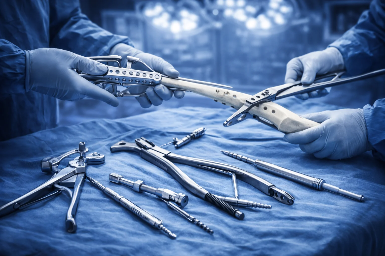
Provides matched drill bits and taps for the trauma cannulated screw system used in fixation of femoral neck, ankle, and wrist fractures. Each drill diameter corresponds to a tap that cuts threads in dense cortical bone before lag screw insertion. The complete set ensures hole preparation and tapping follow manufacturer specifications for pull-out strength.
Used to palpate the pedicle wall and fracture hematoma cavity during percutaneous fixation of thoracolumbar burst fractures. The straight probe confirms breach-free pedicle trajectory before the guidewire is advanced in MIS spine trauma cases. It also assesses articular step-off in pilon and plateau fractures when direct visualization is limited.
Applies calibrated tightening torque to pedicle screw set screws and locking plate screws during final construct assembly in spinal and periarticular trauma fixation. The torque wrench prevents over-tightening that could strip screw threads in osteoporotic bone. Trauma teams document the torque setting for each implant to ensure reproducible fixation per the surgical technique guide.
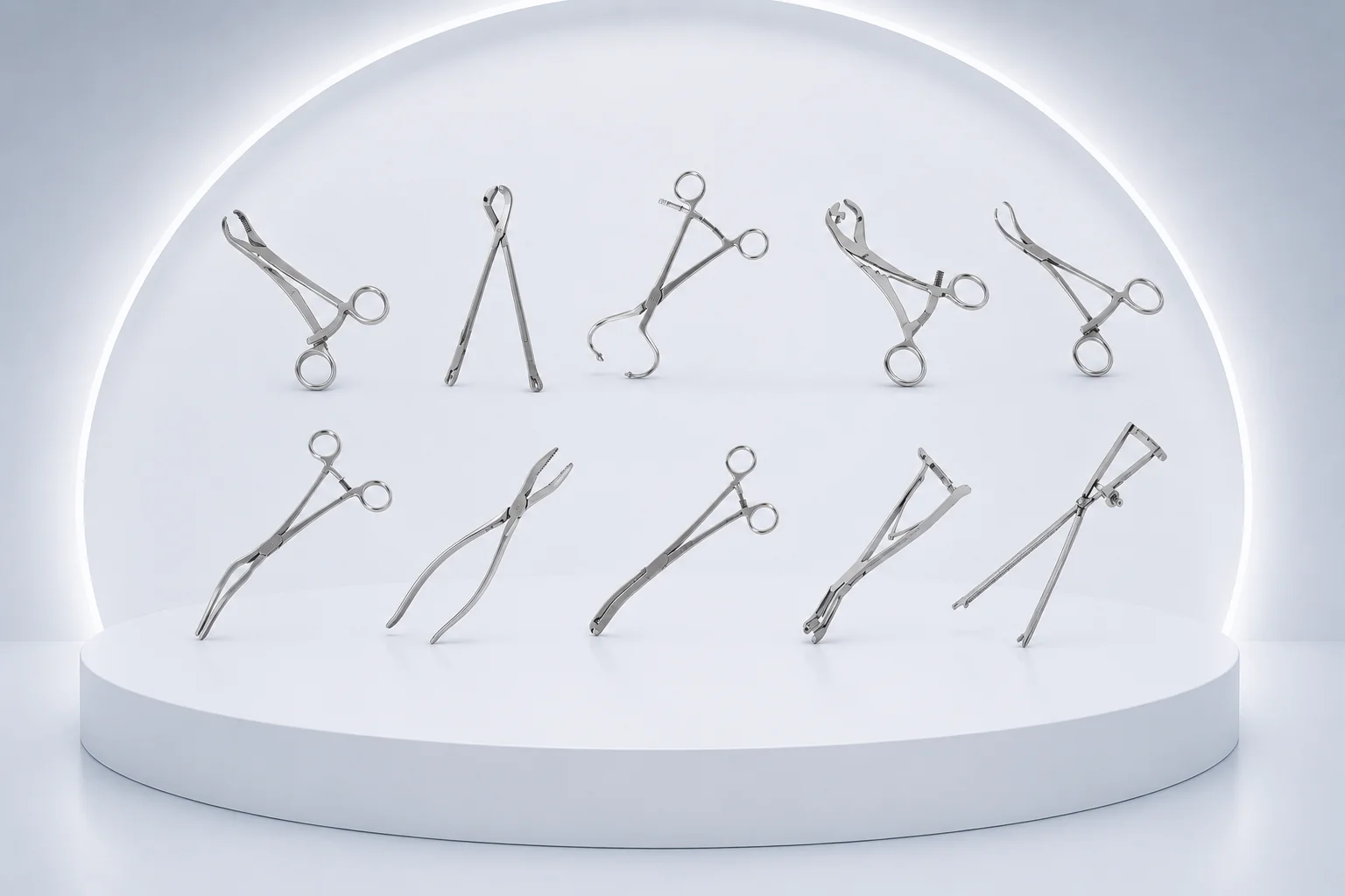
General-purpose trauma reduction forceps for provisional alignment of diaphyseal femur and tibia fractures before intramedullary nailing or plate fixation. The forceps span the fracture gap to restore length and rotation while the surgeon inserts a guidewire or reamer. Emergency trauma teams keep this instrument on the damage-control orthopedics tray for rapid stabilization.
Drives Torx-head locking screws in modern trauma plates and polyaxial pedicle screw systems where hex drivers are insufficient. The sleeved shaft protects surrounding soft tissue from driver slippage during screw insertion through minimally invasive portals. Operating teams use Torx drivers for the final locking step in angular-stable plate systems applied to distal femur fractures.
Drives cannulated lag screws over a guidewire during fixation of femoral neck, scaphoid, and medial malleolus fractures. The hollow shaft threads onto the screw head so torque transfers along the wire axis without displacing the reduction. Trauma surgeons use it when sequential screw insertion is required across a fracture plane under fluoroscopic guidance.